Thursday

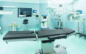
SOPs for an operation theater

 The Standard Operating Procedures (SOPs) for an Operation Theater typically cover various aspects to ensure a sterile and safe environment for surgical procedures. Here's a brief overview:

Pre-Operative Procedures:
  • Verify patient identity, surgical procedure, and consent.
  • Ensure patient fasting requirements are met.
  • Perform pre-operative assessments and screenings.
Operating Room Setup:
  • Sterilize and organize surgical instruments.
  • Confirm availability and functionality of equipment.
  • Ensure cleanliness and aseptic conditions in the operating room.
Personnel Protocols:
  • Verify the credentials of surgical and nursing staff.
  • Follow a strict scrubbing and gowning procedure.
  • Maintain communication protocols during surgery.
Patient Transfer and Positioning:
  • Implement safe patient transfer protocols.
  • Ensure proper patient positioning on the operating table.
  • Confirm proper padding and support for patient comfort.
Aseptic Techniques:
  • Strict hand hygiene and use of sterile gloves.
  • Use of sterile drapes to create a sterile field.
  • Minimize traffic and maintain a closed environment during surgery.
Anesthesia Protocols:
  • Confirm patient's anesthesia history and allergies.
  • Ensure proper administration and monitoring of anesthesia.
  • Follow protocols for airway management and ventilation.
Surgical Procedure Guidelines:
  • Confirm the correct surgical site and procedure.
  • Follow a step-by-step checklist for the surgical process.
  • Document any unexpected events or deviations.
Post-Operative Care:
  • Transfer the patient to the recovery area.
  • Monitor vital signs and assess the patient's condition.
  • Provide post-operative instructions and care.
Cleaning and Sterilization:
  • Dispose of surgical waste appropriately.
  • Follow protocols for cleaning and sterilizing instruments.
  • Maintain a clean and organized operating room after each procedure.
Emergency Response:
  • Establish protocols for handling emergencies.
  • Ensure availability of emergency equipment and medications.
  • Conduct regular drills for the response team.
  • These SOPs are critical to maintaining a high standard of care, minimizing the risk of infections, and ensuring the safety of both patients and healthcare professionals in the operating theater. Specific protocols may vary by healthcare institution and surgical specialties.

Wednesday

Treatment options with their prons and cons


Treatment options with their prons and cons


 Biomedical waste treatment methods aim to reduce the hazard posed by medical waste, ensuring it is safe for disposal or further handling. Here are the common treatment options, along with their advantages and limitations:

1. Autoclaving (Steam Sterilization)

An autoclave is a machine used to carry out  processes requiring elevated temperature and pressure in relation to ambient pressure and/or temperature.

Advantages:

  • Effective at sterilizing infectious waste.
  • Environmentally friendly; uses steam and heat, no harmful emissions.
  • Can be used on a variety of waste types, including plastics and textiles.

Limitations:

  •  Not suitable for all types of waste (e.g., chemical or pharmaceutical waste).
  •  Requires significant energy for heating.
  •  Treated waste may still require shredding for volume reduction.

 2. Incineration

Incineration is the process of burning hazardous materials at temperatures high enough to destroy contaminants. Incineration is conducted in an “incinerator,” which is a type of furnace designed for burning hazardous materials in a combustion chamber. Many different types of hazardous materials can be treated by incineration, including soil, sludge, liquids, and gases.
Advantages:

  • Reduces waste volume significantly (up to 90%).
  •  Destroys pathogens completely.
  • Suitable for all types of biomedical waste, including pharmaceuticals and chemicals.

Limitations:

  • Produces emissions that can include harmful pollutants (requires effective emission control systems).
  • High operational and maintenance costs.
  • Ash residue requires proper disposal.

 3. Chemical Disinfection

Chemical disinfection consists of adding a disinfectant (generally a strong oxidant) to the water, which reacts with the organic matter and microbial organisms. Most frequent chemical disinfection compounds are chlorine dioxide, chlorine, and chloramines on one hand and ozone on the other hand.

Advantages:

  • Effective for liquid waste and certain solid waste types.
  • Can be done on-site with relatively simple equipment.
  • Neutralizes pathogens quickly.

Limitations:

  • Ineffective for waste types that are not easily penetrated by chemicals (e.g., bulk or solid waste).
  • Requires handling and storage of hazardous chemicals.
  • Chemical residues may pose additional disposal challenges.

 4. Microwave Irradiation

It basically works like a souped-up version of your kitchen’s microwave oven. Typical operation is at 2450 Hz. While an autoclave provides heat from outside the waste, like a conventional kitchen oven, the microwave unit transmits energy as microwaves and that energy turns into heat inside the wet waste.

Microwave disinfection works only when there is water in the waste. Because the radiation directly works on the water, not the solid components of the waste. For this reason, treatment units are often supplied with a humidifier. Processing time is determined by the manufacturer and experience of the operators, but somewhere about 20 minutes per batch is typical

Advantages:

  • Effective for disinfecting infectious waste.
  • Reduces the volume of waste.
  • Can be done on-site, reducing transportation risks.

Limitations:

  • Requires shredding of waste prior to treatment, which can be an additional cost and process.
  • Limited capacity; not suitable for all waste types (e.g., large items or chemical waste).
  • High initial setup costs.


 5. Land Disposal (Sanitary Landfills)

In the landfill method of waste disposal, a huge pit is made in an open low lying area, usually away from the places where people reside. The wastes is collected in huge trucks and dumped into the pits. Once the pits are full, they are covered with soil and left for decomposition.

Advantages:

  • Low cost compared to other treatment methods.
  • Simple and straightforward disposal method for treated waste.

Limitations:

  • Risk of groundwater contamination if not properly managed.
  • Requires large land areas.
  • Not suitable for untreated infectious or hazardous waste.

6. Plasma Pyrolysis

It is an environment-friendly technology, which converts organic waste into commercially useful by-products.a process of thermal degradation of the waste in the total absence of air that produces recyclable products, including char, oil/wax and combustible gases. Pyrolysis has been used to produce charcoal from biomass for thousands of years.

Advantages:

  • High-temperature treatment that converts waste into syngas and slag, both of which can be safely disposed of or reused.
  • Produces minimal emissions.
  • Can treat a wide variety of waste types, including hazardous waste.

Limitations:

  • Very high energy consumption.
  • High operational and maintenance costs.
  • Requires sophisticated technology and skilled operators.


 7. Encapsulation

Encapsulation means coating the waste with inert materials. The coating materials are chemically stable, adhere to the waste, and resist biodegradation. High-density polyethylene (HDPE) and polybutadiene are most often used to perform encapsulation.

Advantages:

  • Effective for sharp and highly infectious waste.
  • Simple and relatively low-cost method.

Limitations:

  • Not a standalone method; typically used for final disposal after other treatments.
  • Encapsulation materials must be managed properly to avoid environmental contamination.

8. Thermal Inactivation

Temperature-based inactivation of pathogens (viruses) is the subject of active scientific research. High temperatures can lead to thermal destruction of the constituent parts of the virions, for instance, thermal denaturation of the surface proteins.

Advantages:

  • Effective for treating certain types of biomedical waste (e.g., laboratory waste).
  • Can be integrated into existing heat-treatment processes.

Limitations:

  • Limited to specific waste types.
  • Requires significant energy input.
  • Not effective for all pathogens and waste forms.

Each treatment option has its specific use cases, and often, a combination of methods is employed to handle different types of biomedical waste efficiently and safely. The choice of treatment depends on factors like the type of waste, volume, regulatory requirements, and available resources.

Saturday

Steps of Biomedical waste management cycle in a hospital

 The biomedical waste management cycle in a hospital involves several stages, ensuring that all medical waste is handled safely and disposed of properly. Here's an overview of the typical cycle:


1. Segregation:

   - Waste is segregated at the point of generation.

   - Different types of biomedical waste (e.g., sharps, infectious waste, pathological waste) are separated into color-coded containers according to regulatory guidelines.


2. Collection:

   - Segregated waste is collected in designated bins.

   - Sharps containers, infectious waste bags, and other specific containers are used to ensure safety and compliance.


3. Storage:

   - Collected waste is temporarily stored in a secure, designated area within the hospital.

   - Storage areas are designed to prevent access by unauthorized persons and to minimize the risk of exposure or contamination.


4. Transportation:

   - Waste is transported from the storage area to the treatment or disposal site.

   - Internal transport within the hospital follows strict protocols to prevent spills or exposure.

   - External transport is carried out by licensed biomedical waste handlers following regulatory standards.


5. Treatment:

   - Waste is treated to reduce its hazard potential.

   - Common treatment methods include autoclaving (steam sterilization), incineration, chemical disinfection, and microwaving.

   - The choice of treatment depends on the type of waste and local regulations.


6. Disposal:

   - Treated waste is disposed of in an environmentally safe manner.

   - Incinerated waste results in ash, which is disposed of in landfills.

   - Autoclaved waste may be disposed of as regular waste if it meets safety standards.


7. Record Keeping and Monitoring:

   - Detailed records are kept at each stage of the waste management process.

   - Monitoring ensures compliance with regulations and helps in the continuous improvement of waste management practices.


8. Training and Awareness:

   - Hospital staff receive regular training on biomedical waste management practices.

   - Awareness programs help reinforce the importance of proper waste handling and segregation.


9. Compliance and Audits:

   - Regular audits and inspections are conducted to ensure compliance with local, national, and international regulations.

   - Corrective actions are taken if any non-compliance is identified.


Biomedical waste management cycle in a hospital


Implementing an effective biomedical waste management cycle helps protect hospital staff, patients, and the environment from the risks associated with biomedical waste.

Wednesday

How to check & calibrate anesthesia equipment ?

 Anesthesia equipment checking and calibration are crucial steps in ensuring the safety and effectiveness of medical procedures in the operating theater. 


Here's a step-by-step explanation:


Preparation:


Gather the necessary tools and documentation, including calibration certificates, user manuals, and any specific checklists provided by the equipment manufacturer.


Visual Inspection:


Examine the anesthesia workstation for any visible damage, loose connections, or signs of wear. This includes checking hoses, cables, and the physical condition of the equipment.


Power-Up Test:


Turn on the anesthesia machine and associated equipment to ensure proper power-up sequences. Verify that all components, such as monitors and ventilators, initiate without errors.


System Check:


Perform a system self-test if available. This test helps identify any internal malfunctions within the anesthesia machine.


Gas Supply Verification:


Confirm the availability of medical gases and check the pressure levels. Ensure that the pressure regulators are functioning correctly.


Ventilator Calibration:


Calibrate the ventilator to ensure accurate tidal volume delivery, respiratory rate, and other parameters. Use a calibrated test lung to verify the ventilator's performance.


Gas Flow and Vaporizer Calibration:


Calibrate the flow meters to guarantee accurate gas delivery rates. Check and calibrate vaporizers for volatile anesthetics to ensure precise administration.


Oxygen Analyzers:


Calibrate and verify the accuracy of oxygen analyzers. Confirm that the concentration of inspired oxygen is within the specified range.


Pressure Monitoring:


Calibrate pressure monitoring devices, including those for airway pressure, PEEP (positive end-expiratory pressure), and other relevant parameters.


Temperature and Humidity Control:


Ensure that the temperature and humidity control systems, if present, are functioning properly to maintain a suitable environment for the equipment.


Alarms Testing:


Test and verify the proper functioning of alarms, including high and low pressure, low oxygen concentration, and any other alarms specified by the equipment.


Documentation:


Record the results of the checks and calibrations in a log or checklist. Include details such as date, time, equipment condition, and any corrective actions taken.


Regular Maintenance:


Establish a routine maintenance schedule for the anesthesia equipment based on the manufacturer's recommendations. Regularly check and update calibration records.


Staff Training:


Ensure that the healthcare personnel operating the anesthesia equipment are trained on proper usage, checks, and emergency procedures.


Regular and thorough anesthesia equipment checking and calibration are essential to maintain patient safety and the overall effectiveness of medical procedures in the operating theater. It also helps in compliance with regulatory standards and ensures that the equipment functions as intended.

Sunday

Emergency protocols in a main operating theater

Emergency protocols in a main operating theater are crucial for ensuring the safety and well-being of both patients and medical staff.

 Here's a step-by-step overview:


  • Recognition of Emergency:

Promptly identify the emergency situation.

Common emergencies include cardiac arrest, respiratory distress, massive bleeding, or anesthetic complications 

  • Initiate Emergency Response:

Activate the emergency response system, typically by calling for a "Code Blue" or equivalent

Emergency protocols in a main operating theater


Ensure communication with all necessary personnel, including anesthesia, nursing, and surgical teams.


  • Assessment and Stabilization:


Evaluate the patient's vital signs and clinical status.

Begin immediate interventions to stabilize the patient, such as basic life support (BLS) or advanced cardiac life support (ACLS) measures.


  • Team Coordination:


Assign specific roles to team members.

Encourage effective communication and teamwork among the surgical, anesthesia, and nursing staff.


  • Anesthesia Considerations:


If the emergency is related to anesthesia, follow specific protocols for managing complications.

Adjust anesthesia levels as needed, and be prepared to secure the airway.


  • Surgical Intervention:


Consider the necessity of halting the surgical procedure.

Prioritize life-saving interventions over the ongoing surgical procedure.


  • Communication with Family:


Designate a team member to communicate with the patient's family.

Provide updates on the situation and address their concerns with empathy.

Equipment and Medication Availability:

Ensure that all necessary emergency equipment and medications are readily available.

Regularly check and maintain the functionality of emergency equipment.


  • Documentation:


Document the events, interventions, and responses during the emergency.

This documentation is crucial for subsequent analysis and quality improvement.


  • Debriefing and Review:


Conduct a debriefing session after the emergency situation is resolved.

Analyze the events, identify areas for improvement, and update protocols as needed.

Tuesday

Inspection and Maintenance Sterilization Procedures in Operation Theater

 Inspection and Maintenance Sterilization Procedures in Operation Theater:

1.Preparation:

Ensure all necessary tools and equipment are gathered.

Verify the availability of sterilization agents and solutions.

2.Initial Inspection:


Examine the sterilization equipment for visible damage or wear.

Check seals, gaskets, and valves for integrity.

Inspect trays, containers, and packaging materials for defects.

3.Functional Testing:

Conduct functional tests on sterilizers, ensuring proper functioning of controls.

Test pressure gauges, temperature indicators, and safety mechanisms.

Inspection and Maintenance Sterilization Procedures in Operation Theater:


4. Cleaning and Decontamination:

Thoroughly clean and decontaminate all instruments and equipment.

Follow established protocols for cleaning to remove organic material.

5. Assembly Check:

Ensure that instruments are correctly assembled and arranged for effective sterilization.

Check compatibility of materials with chosen sterilization methods.

6. Packaging:

Employ proper packaging techniques to maintain sterility.

Use appropriate indicators to confirm successful sterilization.

7. Loading:

Load sterilization chambers according to equipment specifications.

Avoid overloading to allow for proper circulation of sterilizing agents.

8. Sterilization Process:

Initiate the sterilization process adhering to recommended parameters.

Monitor and record sterilization time, temperature, and pressure.

9. Post-Sterilization Inspection:

After sterilization, inspect packaging for integrity.

Confirm color changes in indicator strips to ensure effectiveness.

10. Storage:

Store sterilized items in designated areas with controlled environmental conditions.

Follow a first-in, first-out (FIFO) system for inventory management.


11. Documentation:

Maintain detailed records of each sterilization cycle.

Document equipment checks, sterilization parameters, and any anomalies.

12. Regular Maintenance:

Schedule routine maintenance for sterilization equipment.

Address any issues promptly to prevent disruptions in operation.


Training and Compliance:

Ensure staff is adequately trained on sterilization procedures.

Adhere to regulatory guidelines and standards governing sterilization in healthcare.

These procedures are essential for maintaining a sterile environment in the operation theater, reducing the risk of infections and ensuring patient safety.

Wednesday

How to remove bubbles from suctioning tube?

Suctioning can help maintain and establish the gas exchange, adequate oxygenation, and alveolar ventilation. Suctioning can be performed through an endotracheal tube, a tracheostomy tube, the mouth, or the nose.

Suctioning can help maintain and establish the gas exchange, adequate oxygenation, and alveolar ventilation. Suctioning can be performed through an endotracheal tube, a tracheostomy tube, the mouth, or the nose.

How to remove bubbles from suctioning tube or refill a suctioning tube? 

Refilling a suctioning tube typically involves the following steps:

1. Prepare the equipment : Ensure you have a clean suctioning tube and a container of sterile saline solution.


2. Disconnect the tube: If the suctioning tube is connected to any equipment or devices, disconnect it to make the refilling process easier.


3. Fill the container : Fill a sterile container with the desired amount of saline solution. Ensure the container is clean and free from any contaminants.


4. Attach the tube to the container: Attach the end of the suctioning tube to the container of saline solution. Ensure it is securely attached to prevent leaks.


5. Prime the tube : Hold the suctioning tube and container of saline solution upright. Gently squeeze the container to allow the saline solution to flow through the tube and remove any air bubbles. Continue squeezing until the tube is filled with saline solution.


6. Test the suction: Once the tube is filled, test the suction to ensure it is working properly. You can do this by connecting the tube to the suction device and activating it briefly to ensure proper suctioning.


7. Secure connections: Once you've confirmed that the suctioning tube is filled and working correctly, securely reconnect it to any equipment or devices as needed.


8. Dispose of any excess saline solution: Dispose of any remaining saline solution in the container properly according to medical waste disposal guidelines.


9. Clean up: Clean any spills or drips, and ensure all equipment is properly stored.


10. Document: Finally, document the procedure according to your facility's protocols.


Always follow your healthcare facility's specific guidelines and protocols for refilling suctioning tubes to ensure patient safety and compliance with best practices.

Saturday

Surgical wound classification

Surgical wound classification is typically based on the level of contamination. The categories include:
operationtheatertips.blogspot.com


  • Centers for Disease Control and Prevention (CDC) surgical wound classification

    • class I - clean

      • uninfected operative wound with no inflammation
      • no entry into the respiratory, alimentary, genital, or urinary tract
      • primarily closed, and drained with closed drainage (if necessary)
      • includes operative incisional wounds following blunt trauma if above criteria met

    • class II - clean-contaminated

      • entry into respiratory, alimentary, genital, or urinary tract under controlled conditions without unusual contamination
      • includes operations involving biliary tract, appendix, vagina, and oropharynx if no evidence of infection or major break in technique

    • class III - contaminated

      • fresh, open, accidental wounds
      • operations with major breaks in sterile technique or gross spillage from gastrointestinal tracts 
      • incisions with acute, nonpurulent
      •  inflammation.
      • operationtheatertips.blogspot.com

    • class IV - dirty/infected

      • old traumatic wounds with retained devitalized tissue
      • wounds involving existing clinical infection or perforated viscera
      • suggests infective organisms present in operative field before operation.
    • Reference - CDC Hospital Infection Control Practices Advisory Committee guideline on prevention of surgical infection (Infect Control Hosp Epidemiol 1999 Apr;20(4):250), commentary can be found in Infect Control Hosp Epidemiol 1999 Apr;20(4):231

Tuesday

Processing of laproacpoic instrumemts

 How to clean , disinfect and sterilize Laproscopic instruments?              

Glutaraldehyde formulations are the most popular chemical germicides for high-level disinfection of laparoscopic and endoscopic equipment.  

Processing of laproacpoic instrumemts


Optimal processing of LI involves several steps that reduce the risk of transmitting infection. These are :

1) Dismantling 

Dismantal all removable parts for thorough cleaning. 

2)Decontamination

 The procedure begins in the theatre itself using the nursing staff wiping off visible blood tissue and body fluids in the instruments with a damp sterile sponge. At the conclusion of this all soiled or contaminated instruments should be placed in a container containing a disinfectant solution such as 0.5% chlorine and allowing them to soak for Ten minutes.

3) Precleaning

 Following the instruments reach the sterile supplies processing area, which is preferably a controlled environment, a pre-cleaning treatment with an enzymatic method ( protease, amylase, lipase etc.) is recommended

 4) Cleaning

 Ultrasonic cleaning is 16 times better than hand-cleaning. The instruments are placed in the ultrasonic unit for 10-15 minutes and use a neutral pH solution.

5) Drying

 This really is ideally achieved by using an air gun that blows all the water droplets off the surfaces of instruments or by using an oven. 

6) Sterilization

 There are three sterilization processes available to us - steam, ethylene oxide and glutaraldehyde. 

Flash / vacuum steam sterilization

Laparoscopes may be sterilized by flash or vacuum steam sterilization. Before sterilization, all instruments that are insulated, all silicone tubing, and all sorts of cords ought to be doubly covered with a cloth to prevent connection with the hot metallic container. They are then put into the autoclave. Flash sterilization is carried out at 135 0C at 30 psi pressure for 60 minutes.This method requires post-vacuum and dry cycles. The instruments should rest on a sterilizer rack for 45 minutes to prevent water condensation about the lens.

Gas sterilization

Endoscopic instruments may be sterilized with either cold or warm EO gas, with respect to the manufacturer’s instructions. With cold gas, the temperatures are set at 85 °C and also the instruments are subjected for 4 hours and 30 minutes. Aeration must then follow for 12 hours. Warm gas sterilization happens at 145 °C for 2 hour 30 minutes, followed by 8 hours aeration. The benefits of EO are how the items aren't damaged, it's non-corrosive to optics also it permeates porous material. Its main disadvantages are its cost, toxicity, the requirement for aeration and being a longer process.

High level  disinfection

Agents that are employed for HLD include 2% glutaraldehyde, 6% stabilized hydrogen peroxide and per acetic acid . Glutaraldehyde has got the benefits of having good biocidal activity, non-corrosive to optics and it is active in the presence of protein. Fibreoptic light cords and telescopes have to be soaked in 2% glutaraldehyde not less than Ten minutes. Soaking should not exceed Twenty minutes. The endocamera could also disinfected by 10 minutes submersion in 2% glutaraldehyde. 

7) Storage:

 For optimal storage, sterile packs are put in closed cabinets in areas that aren't heavily trafficked, have moderate temperatures, and are dry or of low humidity

Thursday

Merit and Demerits of Robotic assisted Surgery

Merit and Demerits of Robotic assisted Surgery

Robotic assisted surgery allow a surgeon at a console to operate remote-controlled robotic arms, which may facilitate the performance of procedures. These robotic systems are designed to enhance the surgeon's precision, control, and dexterity, offering potential benefits to both the surgeon and the patient.

1. Robotic System: The surgeon operates from a console equipped with a 3D visualization system and manipulates the robotic arms using hand and foot controls.

2. Enhanced Precision: Robotic systems provide enhanced precision by filtering any hand tremors of the surgeon's movements. The robotic arms translate the surgeon's hand movements into smaller, more precise movements, reducing the risk of human error.

3. Minimally Invasive: Robot-assisted surgery is typically performed using minimally invasive techniques, such as laparoscopy or thoracoscopy. Minimally invasive procedures involve smaller incisions compared to traditional open surgery, resulting in reduced scarring, less pain, and faster recovery for the patient.

4. 3D Visualization: Surgeons using robotic systems benefit from high-definition, 3D visualization of the surgical site. This allows for better depth perception and visualization of tiny anatomical structures, improving surgical accuracy.

5. Telemanipulation: Robotic-assisted surgery can enable telemanipulation, where a surgeon can perform surgery remotely. This has the potential to bring expert surgical care to underserved areas or allow surgeons to operate on patients in different locations without needing to be physically present.

6. Complex Procedures: Robot-assisted surgery is commonly used for complex procedures in various medical specialties, including urology, gynecology, general surgery, thoracic surgery, and colorectal surgery. It has been particularly beneficial in procedures such as prostatectomies, hysterectomies, and colorectal resections.

7. Limitations: While robot-assisted surgery offers many advantages, it also has limitations. The cost of robotic systems can be high, which can limit their accessibility in some healthcare settings. Additionally, the learning curve for surgeons to become proficient in robotic surgery can be steep.

Technical limitations: Robotic surgery relies on precise movements and feedback from the robotic system. However, there may be limitations in certain procedures that require a high degree of tactile feedback or complex maneuvering. The lack of haptic feedback in robotic systems can be a disadvantage in delicate surgical procedures

Dependency on technology: Robot-assisted surgery heavily relies on technology, including robotic systems, software, and infrastructure. In the event of a technical glitch or system failure, the surgery may be interrupted, leading to potential complications and the need to switch to a traditional surgical approach. This dependency on technology can introduce an additional layer of risk and complexity.

 Increased procedure time: Initially, robot-assisted surgeries often take longer to perform compared to traditional procedures. The setup and docking of the robotic system, as well as the need for precise instrument manipulation, can extend the overall duration of the surgery. Prolonged procedure times may increase the patient's exposure to anesthesia, increase the risk of complications, and strain healthcare resources.

 It's important to note that while these demerits exist, the field of robot-assisted surgery continues to evolve, and ongoing advancements are aimed at addressing these limitations and improving patient outcomes.

Sunday

DOSIMETERS

Dosimeters

A batch dosimeter is a type of radiation dosimeter that is designed to measure the amount of radiation exposure over a specific period, typically over several weeks or months. Batch dosimeters are often used in occupational settings where workers may be exposed to radiation regularly, such as in nuclear power plants, medical facilities, or research laboratories.

Batch dosimeters work by using a material that is sensitive to radiation, such as a film or a thermo-luminescent material, which is then exposed to the radiation. After a certain period, the dosimeter is removed from the exposure area and the level of radiation exposure is calculated by measuring the change in the sensitivity of the dosimeter material.


They are also relatively inexpensive and easy to use, making them a practical option for many occupational settings.
Radiation workers who are issued single badges for monitoring whole-body dose should wear them in the region of the collar with the label facing out. When a lead apron is worn, the dosimeter should be outside the lead apron. Technologists who work with fluoroscopy may wear two badges, one on the collar outside the lead apron and one at the waist that is under the apron. The two dosimeters should be distinguished by color or icons indicating their specific locations. Personnel who are issued dosimeters should wear them at all times when working in radiation areas and should keep them in a safe place, away from radiation and heat, when off duty. In addition to whole-body badges, ring dosimeters may be worn by nuclear medicine technologists and others whose work results in more exposure to the hands than to the body.

There are several types of batch dosimeters available, each with its own advantages and disadvantages. Some common types include:

Film badge dosimeters: These dosimeters use a small piece of film that is sensitive to radiation exposure. The film is exposed to radiation over a certain period and then developed to show the level of exposure. Film badge dosimeters are inexpensive and easy to use, but they can be affected by temperature and humidity.

They are still in use today but are much less common. The disadvantage of this type of personal monitor is that the dental film is subject to fog when exposed to heat or fumes, and this exposure could result in a false reading. The film is also ruined if it is laundered! After a period of use, the film is returned to a laboratory that processes it and measures the OD of the film. The exposure is calculated and reported based on this measurement. Many radiographers still refer to their dosimeters as "film badges," but today they are more likely to be TLDs or OSLs.
Except OSL badges, dosimeters cannot accurately measure total exposures of less than 5 mrem (0.05 mSv). For this reason, personnel who receive very small amounts of exposure will get more accurate measurements with less frequent badge changes. Personnel involved in diagnostic radiography who are always or nearly always in a control booth during exposures are usually best monitored with quarterly service. Monthly service is a better choice for those who work in fluoroscopy and those who perform bedside radiography.

Service companies provide an extra dosimeter in every batch that is marked "CONTROL." The purpose of this dosimeter is to measure any radiation exposure to the entire batch while in transit. Any amount of exposure measured from the control badge will be subtracted from the amounts measured from the other badges in the batch. The control badge should be kept in a safe place, away from any possibility of x-ray exposure. It should never be used to measure occupational dose or for any other purpose.
Radiation badge service companies will want to know the name, birth date, and Social Security number of all persons to be monitored so that all records can be accurately identified. If there has been a history of previous occupational radiation exposure and the dose is known, this information should also be provided so that the record will be complete and accurate. Exposure reports are sent to the subscriber for each batch, and an annual summary of personal exposure is also provided. Radiation workers should be advised of the radiation exposure reported from their badges and should be provided with copies of the annual reports for their own records. Employees exposed to ionizing radiation should not leave their employment without a complete record of their radiation exposure history. Employers are required to provide this information.


Thermo-luminescent dosimeters (TLDs): TLD stands for thermoluminescent dosimeter. The roots of this term mean "dose-measuring device that gives off light when heated." The TLD is a type of personal monitor commonly used by radiographers.
It consists of a plastic badge or ring containing one or more lithium fluoride crystals. These crystals (and several others with similar characteristics) absorb x-ray energy and, when heated, give off the energy again in the form of light. The TLD is more durable than the film badge insert and responds only to ionizing radiation exposure. TLDs are more accurate than film badge dosimeters, but they are also more expensive.


Optically Stimulated Luminescence (OSL) dosimeters: These dosimeters use a small piece of aluminum oxide that is sensitive to radiation exposure. When the crystal is exposed to radiation, it traps the energy from the radiation. The dosimeter is then read using a laser that causes the crystal to emit light, and the amount of light emitted is proportional to the amount of radiation exposure. OSL dosimeters are very accurate and can be reused, but they are also more expensive than other types of dosimeters.

Pocket ionization chambers: These dosimeters use a small chamber filled with air that is sensitive to radiation exposure. When radiation passes through the chamber, it ionizes the air, which can be measured to determine the level of exposure. Pocket ionization chambers are very accurate, but they are also expensive and require regular calibration.


The choice of dosimeter type will depend on the specific needs of the user and the type of radiation exposure monitored.

Research :

We present the characteristics of a new silicone-based radio chromic dose-response dosimeter containing the leuco-malachite green (LMG) dye. The dose-response as well as the dose rate and photon-energy dependence of the dosimeter were characterized. To optimize the dose-response, different concentrations of the chemical components were investigated. The dose-response was found to decrease exponentially as a function of time after irradiation. A cylindrical dosimeter was produced and irradiated with a volumetric modulated arc therapy plan; the standard deviation between the measured and calculated dose was 5% of the total dose.

A new dosimeter formulation for deformable 3D dose verification

 and 

Published under license by IOP Publishing Ltd

Featured Post

How to manage hospital waste? HOSPITAL WASTE :  Hospital waste is “Any waste which is generated in the diagnosis, treatment or immunization ...