Sunday

Tips to prevent fogging on glasses when wearing a mask

Tips to prevent fogging on glasses when wearing a mask

Tips to prevent fogging on glasses when wearing a mask 

 Here are some tips to prevent fogging on glasses when wearing a mask:

The standard technique of tying a surgical mask involves knotting the two ties so they lie above and below the ear in a near parallel appearance (Fig 1). 
Tips to prevent fogging on glasses when wearing a mask

Our method consists of knotting the superior tie first with it lying directly below the ear. The inferior tie is brought up in front of the ear and knotted over the crown of the head (Fig 2).
Tips to prevent fogging on glasses when wearing a mask


  1. Clean your glasses with soap and water before putting on your mask.
  2. Adjust your mask to fit securely and tight on your face.
  3. Create a seal around your nose by pressing down on the top of the maskTips to prevent fogging on glasses when wearing a mask
  4. Breathe out slowly and calmly.
  5. Try using a mask with a moldable nose bridge or insert a small piece of tissue under the top of the mask.
  6. Wear glasses with anti-fog coating.

Tying a surgical mask to prevent fogging
Authors: DJ Jordan and R Pritchard-JonesAUTHORS INFO & AFFILIATIONS
Tuesday

Retractor Fatigue During Surgery

 Retractor Fatigue During Surgery

A retractor is a surgical instrument used to hold back or separate tissues during a surgical procedure. It is typically a flat or curved blade with a handle that can be adjusted to various angles. Retractors are commonly used in various types of surgeries, such as abdominal, thoracic, and orthopedic procedures, to provide better visibility and access to the surgical site. They can also be used to hold organs or other structures in place during a procedure.

Retractor Fatigue During Surgery


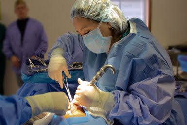
Retractor fatigue during surgery refers to the physical exhaustion that can occur in surgeons when using retractors for prolonged periods of time during an operation. This can lead to decreased precision and increased risk of errors. Manual retraction, a task performed to expose the surgical site, poses a high risk for musculoskeletal disorders that affect the hands, arms, shoulders, neck, and back(1).  To prevent retractor fatigue, surgeons may take frequent breaks, use assistive devices, or switch to minimally invasive techniques when possible.

Correct way of retraction:

  • Start by selecting the appropriate retractor for the surgical procedure.
    Retractor Fatigue During Surgery

  • Position the retractor at the surgical site before making incisions.
  • Use gentle pressure to hold back or separate the tissues.
  • Adjust the angle of the retractor as needed to provide optimal visibility and access to the surgical site.
  •  Be mindful of the surrounding structures and avoid applying excessive force or pressure, as this can cause damage to surrounding tissues.
  • Keep the retractor in place throughout the procedure, and adjust as necessary to ensure proper visibility and access.
  • Once the procedure is complete, remove the retractor and close the incision site.

It's essential to use the appropriate retractor for the procedure, be gentle and avoid applying excessive force or pressure, and keep the retractor in place throughout the procedure and adjust as necessary to ensure proper visibility and access.  

1.AORN Ergonomic Tool 5: Tissue Retraction in the Perioperative SettingAuthor links open overlay panel

Wednesday

What is suctioning and its effects?

What is suctioning and its  effects?

Suctioning of fluid during surgery is a common practice that helps to maintain a clear and safe surgical field. It is typically done using a suction device, such as a surgical suction device, which is a tube connected to a source of suction, such as a vacuum.

Fluid suctioning during surgery can be used to remove blood, saliva, and other bodily fluids that may accumulate in the surgical field. This is important for maintaining visibility for the surgeon and preventing fluid from entering the patient's lungs  and causing complications. Suctioning can also be used to remove debris, such as bone chips, during orthopedic procedures.

What is suctioning and its  effects?


During surgery, the surgical team may use various types of suction catheters to remove fluid and debris. These can include rigid suction catheters, which are used to clear large amounts of fluid, and soft suction catheters, which are used to remove smaller amounts of fluid. Additionally, suction may be used during surgery to clear the airway of a patient.

What complications can happen due to suctioning?

Suctioning can also cause trauma to the tissue, which can lead to bleeding, inflammation, and other complications. It can also cause hypotension due to decreased blood flow and decreased blood pressure, and it can also cause bronchospasm and laryngeal spasms. In addition, suctioning can also lead to hypoxemia, which is a decrease in the amount of oxygen in the blood.

 How much time suctioning can be used?

The duration of suctioning depends on the individual case and the specific condition being treated. In general, suctioning should only be used for a short period , usually no more than 10-15 minutes at a time. It is important to consult with a healthcare professional for guidance on how long suctioning should be used for your specific situation. Additionally, it's important to take breaks in between suctioning sessions, and not to use suctioning excessively as it can cause irritation or even injury to the airway.


Saturday

Surgical site infection, its types & Prevention

Surgical site infection, its types &  Prevention

An infection that develops at the site of surgery is known as a surgical site infection (SSI). SSIs can happen up to a year after some procedures, like knee replacements, or within 30 days following an operation. They can be minor to severe, cause healing to be delayed, or possibly result in sepsis or death.

Surgical site infections (SSIs) are a common complication of surgery, affecting up to 5% of patients who undergo invasive procedures. SSI  risk factors include poor hygiene, diabetes, obesity, smoking, and certain types of surgery. SSIs can be prevented by following proper surgical techniques, using appropriate antibiotics, and maintaining good hygiene before and after surgery.

Surgical site infection, its types &  Prevention

Types Of SSI

  • Superficial Incisional SSI

 Infection occurs within 30 days of the surgical procedure. Infection involves only skin or subcutaneous tissue of the incision and at least one of the following symptoms from the superficial incision unless the wound is culture negative:

1. Purulent drainage, x-ray with or without laboratory confirmation, from the incision

2. Organisms isolated from an aseptically obtained culture of fluid or tissue

3. At least one of the following infections' warning signs or symptoms:

a. Pain or tenderness                             b. Localized swelling

c. Redness or heat                                 d. Surgeon deliberately opens the incision

4. Diagnosis of superficial incisional SSI by the surgeon or physician

Do not report the following conditions as superficial SSI:

1. Stitch abscess (minimal inflammation and discharge) limited to the points of suture penetration

2. Infection of episiotomy or newborn circumcision site (these have separate criteria)

3. Infected burn wound

4. Incisional SSI that extends into the fascial and muscle layers 

Surgical site infection, its types &  Prevention


  • Deep incisional SSI

Infection occurs within 30 days after the procedure if no implant is left in place or within 1 year if an implant is left in place and the infection appears to be related to the procedure. Infection involves deep soft tissues (e.g., fascia and muscle layers) of the incision and at least one of the following symptoms unless the site is culture negative:

1. Purulent drainage from the deep aspects of the incision, but not the organs or compartments

2. A deep incision spontaneously dehisces or deliberately opened by the surgeon because of:

a. Fever .100.4° F (38° C)

b. Localized pain

3. Diagnosis of deep SSI by physician

or surgeon

Additional factors:

1. Report combined superficial and deep infections as deep SSI

2. Report organ space or deep infection that drains through the incision as  deep SSI

Treatment typically includes antibiotics and may involve further surgical intervention if the infection is severe.

Organ/space surgical site infections (SSIs) are infections that occur within an organ or body cavity after surgery. They can occur when bacteria enter the body during the surgery and infect the surrounding tissues. Organ/space SSIs are considered more serious than superficial or deep incisional SSIs as they can lead to sepsis and potentially death. Risk factors include prolonged surgery, poor wound healing, and certain underlying medical conditions. Treatment typically includes antibiotics and may involve further surgical intervention if the infection is severe.

In the event that a patient does develop an SSI, it is essential to identify the cause and implement appropriate treatment. This may include antibiotics, wound debridement, or in some cases, a return to the operating room for surgical intervention.

Overall, SSIs are a serious complication of surgery that can have significant consequences for patients. However, through proper prevention and management, the risk of infection can be minimized. By understanding the risk factors and implementing appropriate measures, healthcare professionals can help ensure the best possible outcomes.

From The Hospital Infection Control Practices Advisory Committee: Guideline for prevention of surgical site infection, Vol. 20, No. 4, 1999.

Available at: http://www.cdc.gov/ncidod/dhqp/pdf/guidelines/SSI.pdf.

Thursday

What types of drain are most commonly used?

What types of drain are most commonly used?

A surgical drain is a tube used to remove pusblood or other fluids from a wound [1] body cavity, or organ. They are commonly placed by surgeons or interventional radiologists after procedures or some types of injuries, but they can also be used as an intervention for decompression. 

 Drains are classified by various systems: open or closed and passive or active

Passive drains rely on gravity, body movement, pressure differentials, or overflow to move fluid or gas; Penrose drain - a soft rubber tube

Active drains use intermittent or continuous negative pressure to pull fluid or gas from a wound or body cavity.

Jackson-Pratt drain - consists of a perforated round or flat tube connected to a negative pressure collection device. The collection device is typically a bulb with a drainage port which can be opened to remove fluid or air. After compressing the bulb to remove fluid or air, negative pressure is created as the bulb returns to its normal shape.

Negative pressure wound therapy - Involves the use of enclosed foam and a suction device attached; this is one of the newer types of wound healing/drain devices which promotes faster tissue granulation, often used for large surgical/trauma/non-healing wounds.

Blake drain - a round silicone tube with channels that carry fluid to a negative pressure collection device. Drainage is thought to be achieved by capillary action, allowing fluid to travel through the open grooves into a closed cross section, which contains the fluid and allows it to be suctioned through the tube.[2]

Redivac drain - a high negative pressure drain. Suction is applied through the drain to generate a vacuum and draw fluids into a bottle.

Pigtail drain - has an exterior screw to release the internal "pigtail" before it can be removed.

Chest tube - is a flexible plastic tube that is inserted through the chest wall and into the pleural space or mediastinum
  • Wound manager 

What types of drain are most commonly used?

  • References[edit]

    1. ^ Jain, Sudhir Kumar; Stoker, David L.; Tanwar, Raman (2013-04-30). Basic Surgical Skills and Techniques. JP Medical Ltd. pp. 70–73. ISBN 9789350903759. Retrieved 2014-09-07.
    2. ^ Obney, James; Mary Barnes; et al. (2000). "A method for mediastinal drainage after cardiac procedures using small silastic drains". The Annals of Thoracic Surgery70 (3): 1109–110. 10.1016/s0003-4975(00)01800-2. PMID 11016389.

Wednesday

How to manage hospital waste?

How to manage hospital waste?

HOSPITAL WASTE : 

Hospital waste is “Any waste which is generated in the diagnosis, treatment or immunization of human beings or animals or in research” in a hospital.

Hospital Waste Management means the management of waste produced by hospitals using such techniques that will help to check the spread of diseases.

Waste Minimisation Hierarchy 
  • Reduce
  • Reuse 
  • Recycle 
WHO Medical Waste Categories 
  • Infectious
  • Non-Infectious
  •  Hazardous
  • Non Hazardous 
Segregation by color coding system 

Three categories
Infectious waste - Red bags 
Domestic waste - Green Bags 
Sharps -  Needle cutters / Puncture proof containers 
Segregation at Source ( ward, operation theater, laboratory, labor room, othere places).
How to manage hospital waste?


SEGREGATION 

The different waste groups have different colours for the containers and bags for the identification according to the hazards and applied throughout the complete disposal chain (segregation, collection, storage, transport, disposal):
➢ Warning colors for hazardous waste (Red, yellow, orange) 
➢ Positive colors for recycling (Blue, green, etc.)
➢ Neutral colors for normal waste (Black, etc.) 
The colour coding makes the process understandable even for low-skilled workers with language and read problems.

Proper disposal of waste 
 
All infectious waste and sharps containers :Incineration 
All Domestic waste : Landfill 
All hazardous waste : Chemical treatment before disposal 

Hazards associated with poor health care waste management
  • Injuries from sharps to all categories of health care personnel and waste handlers.
  •  Increase risk of infections to medical, nursing and other hospital staff.
  •  Poor infection control can lead to nosocomial infections in patients particularly HIV, Hepatitis B & C.
  •  Increase in risk associated with hazardous chemicals and drugs being handled by persons handling wastes
  •  Poor waste management encourages unscrupulous persons to recycle disposables and disposed drugs for   repacking   and reselling.
  • Organic portion ferments and attracts fly breeding.


Monday

Microbiology of skin

 Microbiology of skin :

The skin is inhabited by the following organisms:

   Transient organisms

• Acquired by direct contact.

• Usually loosely attached to the skin surface.

Mechanically removed by thorough washing.

Resident organism 

Found below the skin surface in hair follicles and sweat glands.

•  Adherent to skin surface 

Chemically removed by using cleaning agent.

Microbiology of skin

Reserch: Skin Preparation for the Prevention of Surgical Site Infection: Which Agent Is Best?

Micah L Hemani, MD and Herbert Lepor, MD

To assess the efficacy of cutaneous antisepsis to prevent catheter-associated infection, we

prospectively studied three antiseptics for disinfection of patients.

The most common agent used today include product containing iodophor or CHG.


Sunday

Why Air Handling system (HVAC) is necessary inside operation theater?

Why Air Handling system (HVAC) is necessary inside operation theater?  

Heating, ventilation, and air conditioning (HVAC) is the use of various technologies to control the temperaturehumidity, and purity of the air in an enclosed space. Its objective is to provide thermal comfort and acceptable indoor air quality.

What are the CDC (central depository company) Guidelines for Isolation Rooms?

  • Air Filtration flow rates to attain 12 air changes per hour (ACH)
  • Negative Pressure to be greater than .01 inches w.g.
  • Monitoring to validate negative pressure

Air Filtration efficiency to be 99.97% or higher as measured against0.3-micronn particle size (HEPA).

Heating, ventilation, and air conditioning (HVAC)


Full Fresh Air: 

Why Air Handling system (HVAC) is necessary inside operation theater?


Importance of HEPA(high efficacy particle air filter) standard:

Here are some specific reasons why ventilation is important in an OT:

Maintaining Sterility: During surgery, the patient's body is exposed, and the surgeon must make incisions and operate on the tissues. The air in the OT can carry bacteria and other contaminants that could infect the open wound. The ventilation system helps to control the airflow and filter out the impurities, reducing the risk of infection.

Reducing Cross-Contamination: Patients undergoing surgery are often susceptible to infections, and the medical staff in the OT must take extra precautions to avoid cross-contamination. Proper ventilation helps to control the flow of air and reduce the spread of airborne particles that can transmit infections.

Removing Odors: Surgery often involves the use of chemicals and other substances that can produce strong odors. The ventilation system helps to remove these odors from the air, making the environment more comfortable for patients and staff.

Controlling Humidity: High humidity levels in the OT can promote the growth of mold and bacteria. Proper ventilation helps to regulate the humidity levels, reducing the risk of infection and creating a more comfortable environment.


Overall, ventilation is a critical component of OT design and operation. It helps to maintain a sterile environment, reduce the risk of infection, and ensure the safety and comfort of patients and medical staff.


Wednesday

How hand washing and scrubbing differentiate?


Hand washing  :

Wash with vigorous mechanical action on all hand surfaces for at least 40 seconds  .

• Rinse under poured or running water. 

To effectively reduce the growth of germs on hands, hand rubbing must be performed by following steps .

• Take only 20 - 30 seconds


Surgical Scrub :

• Process of removing as many microorganisms as possible with the help of scrub brush and &  chemical antiseptic soap .

• Six drops (about 2 to 3 mL) of solution is used• 

A vigorous 2- to 5-minute is effective.

How hand washing and scrubbing differentiate?

Steps for the Brush and Stroke Method:

Gather Supplies: You will need an antimicrobial soap, a sterile brush or sponge (with bristles on one side), and sterile water.

Initial Hand Rinse: Wet your hands and forearms thoroughly under warm running water.

Apply Antimicrobial Soap: Dispense a generous amount of antimicrobial soap onto your hands and forearms.

Scrub Fingernails and Cuticles:

Use the bristled side of the brush to clean under your nails and around your cuticles

Stroke each nail 30 times (recommended in surgical settings) in a downward motion.

Rinse the brush intermittently in sterile water to remove debris.

Scrub Each Finger:

Start with the fingertips and scrub each finger individually, focusing on the spaces between fingers. Use 10–20 strokes for each side of the finger.

Repeat this process for each finger on both hands.

Scrub the Palms and Back of the Hands:

Use the sponge side of the brush to scrub the palms in a circular motion.

Scrub the back of the hands using a similar stroke pattern, paying extra attention to the knuckles and other folds of the skin.

Scrub the Wrists and Forearms:

Move up to the wrists and scrub in circular strokes.

Gradually scrub up to the elbows in circular motions, using long, sweeping strokes, especially in surgical settings.

Cleaning of the entire forearm.

Rinse Thoroughly: Rinse your hands and forearms under sterile running water, keeping your hands higher than your elbows to avoid water running from the elbows back to the hands.

Dry Hands: After scrubbing, dry your hands and forearms with a sterile towel, again ensuring your hands remain elevated above your elbows.

Preparation before hand hygiene :

• The skin and  nails should be kept clean.

• A non oil based product is recommended to protect the skin.

• Fingernails should not reach beyond the fingertips to avoid glove puncture.

• Fingernail polish should not be chipped or cracked.

• All jewelry is removed from the fingers, wrists and neck .

• All hair should be covered by head wear .

• Adjust the disposable mask snugly and comfortably over  the nose and mouth.

 Comfortable water temperature is used as hot water increases chafing and cooler water helps to minimize dry skin.


How to do fumigation in operation theater?

Fumigation in an operating theater is a process of using gas or fumes to kill bacteria, viruses, and other microorganisms that may be present in the air or on surfaces in the operating room. This is done to reduce the risk of infection and ensure that the operating theater is sterile. 

There are several types of fumigants that may be used in the OR, including hydrogen peroxide, ozone, and chlorine dioxide. These fumigants are highly effective at killing microorganisms, and they can be used to decontaminate the air and surfaces in the OR.

Methods of fumigation :

Electric boiler fumigation method : For each 1000 cubic feet of the volume of operation theater 500 ml of formaldehyde(40%solution) added in 1000 ml of water in an electric boiler. Switch on the boiler, leave the room & seal the door . After 45 minutes switch off the boiler without entering in to the room.

Potassium permanganate method : For every 1000 cubic feet add 450 gm of KMnO4 to 500 ml of formaldehyde . Take this mixture in  about 5-8 bowels and allow them to auto boil & generate fumes . After generation of formaldehyde vapor, immediately leave the room & seal it for at least 48 hours.

Here are the steps for fumigating an operating theater:
  • Close all doors and windows in the operating theater, and turn off any ventilation or air conditioning systems.
  • Place fumigation tablets or liquid in a container, such as a vaporizer or fogger. These tablets or liquid will release gas or fumes when activated.
  • Turn on the fumigation device and allow it to run for the recommended amount of time, according to the manufacturer's instructions.
  • After the fumigation process is complete, ventilate the operating theater by opening windows and doors and turning on the ventilation or air conditioning systems.
  • Wipe down all surfaces in the operating theater with a disinfectant solution to remove any residue from the fumigation process.
  • Test the air quality in the operating theater using a gas monitor or other testing device to ensure that it is safe for us.
  • Fumigation frequency depends on:
  • 1. If cases exceeds 15 in 24 hour  
  •   2- Any infectious case     
  •  3- After 1 week
It is important to follow all safety precautions when fumigating an operating theater, as some fumigants can be toxic to humans. Wear protective clothing, such as a mask and gloves, and follow the manufacturer's instructions carefully to ensure the safe and effective use of the fumigant.

How to do fumigation in operation thater?

Featured Post

How to manage hospital waste? HOSPITAL WASTE :  Hospital waste is “Any waste which is generated in the diagnosis, treatment or immunization ...