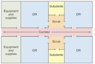
Sunday

DOSIMETERS

Dosimeters

A batch dosimeter is a type of radiation dosimeter that is designed to measure the amount of radiation exposure over a specific period, typically over several weeks or months. Batch dosimeters are often used in occupational settings where workers may be exposed to radiation regularly, such as in nuclear power plants, medical facilities, or research laboratories.

Batch dosimeters work by using a material that is sensitive to radiation, such as a film or a thermo-luminescent material, which is then exposed to the radiation. After a certain period, the dosimeter is removed from the exposure area and the level of radiation exposure is calculated by measuring the change in the sensitivity of the dosimeter material.


They are also relatively inexpensive and easy to use, making them a practical option for many occupational settings.
Radiation workers who are issued single badges for monitoring whole-body dose should wear them in the region of the collar with the label facing out. When a lead apron is worn, the dosimeter should be outside the lead apron. Technologists who work with fluoroscopy may wear two badges, one on the collar outside the lead apron and one at the waist that is under the apron. The two dosimeters should be distinguished by color or icons indicating their specific locations. Personnel who are issued dosimeters should wear them at all times when working in radiation areas and should keep them in a safe place, away from radiation and heat, when off duty. In addition to whole-body badges, ring dosimeters may be worn by nuclear medicine technologists and others whose work results in more exposure to the hands than to the body.

There are several types of batch dosimeters available, each with its own advantages and disadvantages. Some common types include:

Film badge dosimeters: These dosimeters use a small piece of film that is sensitive to radiation exposure. The film is exposed to radiation over a certain period and then developed to show the level of exposure. Film badge dosimeters are inexpensive and easy to use, but they can be affected by temperature and humidity.

They are still in use today but are much less common. The disadvantage of this type of personal monitor is that the dental film is subject to fog when exposed to heat or fumes, and this exposure could result in a false reading. The film is also ruined if it is laundered! After a period of use, the film is returned to a laboratory that processes it and measures the OD of the film. The exposure is calculated and reported based on this measurement. Many radiographers still refer to their dosimeters as "film badges," but today they are more likely to be TLDs or OSLs.
Except OSL badges, dosimeters cannot accurately measure total exposures of less than 5 mrem (0.05 mSv). For this reason, personnel who receive very small amounts of exposure will get more accurate measurements with less frequent badge changes. Personnel involved in diagnostic radiography who are always or nearly always in a control booth during exposures are usually best monitored with quarterly service. Monthly service is a better choice for those who work in fluoroscopy and those who perform bedside radiography.

Service companies provide an extra dosimeter in every batch that is marked "CONTROL." The purpose of this dosimeter is to measure any radiation exposure to the entire batch while in transit. Any amount of exposure measured from the control badge will be subtracted from the amounts measured from the other badges in the batch. The control badge should be kept in a safe place, away from any possibility of x-ray exposure. It should never be used to measure occupational dose or for any other purpose.
Radiation badge service companies will want to know the name, birth date, and Social Security number of all persons to be monitored so that all records can be accurately identified. If there has been a history of previous occupational radiation exposure and the dose is known, this information should also be provided so that the record will be complete and accurate. Exposure reports are sent to the subscriber for each batch, and an annual summary of personal exposure is also provided. Radiation workers should be advised of the radiation exposure reported from their badges and should be provided with copies of the annual reports for their own records. Employees exposed to ionizing radiation should not leave their employment without a complete record of their radiation exposure history. Employers are required to provide this information.


Thermo-luminescent dosimeters (TLDs): TLD stands for thermoluminescent dosimeter. The roots of this term mean "dose-measuring device that gives off light when heated." The TLD is a type of personal monitor commonly used by radiographers.
It consists of a plastic badge or ring containing one or more lithium fluoride crystals. These crystals (and several others with similar characteristics) absorb x-ray energy and, when heated, give off the energy again in the form of light. The TLD is more durable than the film badge insert and responds only to ionizing radiation exposure. TLDs are more accurate than film badge dosimeters, but they are also more expensive.


Optically Stimulated Luminescence (OSL) dosimeters: These dosimeters use a small piece of aluminum oxide that is sensitive to radiation exposure. When the crystal is exposed to radiation, it traps the energy from the radiation. The dosimeter is then read using a laser that causes the crystal to emit light, and the amount of light emitted is proportional to the amount of radiation exposure. OSL dosimeters are very accurate and can be reused, but they are also more expensive than other types of dosimeters.

Pocket ionization chambers: These dosimeters use a small chamber filled with air that is sensitive to radiation exposure. When radiation passes through the chamber, it ionizes the air, which can be measured to determine the level of exposure. Pocket ionization chambers are very accurate, but they are also expensive and require regular calibration.


The choice of dosimeter type will depend on the specific needs of the user and the type of radiation exposure monitored.

Research :

We present the characteristics of a new silicone-based radio chromic dose-response dosimeter containing the leuco-malachite green (LMG) dye. The dose-response as well as the dose rate and photon-energy dependence of the dosimeter were characterized. To optimize the dose-response, different concentrations of the chemical components were investigated. The dose-response was found to decrease exponentially as a function of time after irradiation. A cylindrical dosimeter was produced and irradiated with a volumetric modulated arc therapy plan; the standard deviation between the measured and calculated dose was 5% of the total dose.

A new dosimeter formulation for deformable 3D dose verification

 and 

Published under license by IOP Publishing Ltd

Monday

PHYSICAL LAYOUT OF THE SURGICAL SUITE


PHYSICAL LAYOUT OF THE SURGICAL SUITE 

The physical layout of an operating theater, also known as an operating room, can vary depending on the hospital or surgical facility.

Efficient use of the physical facilities is important. The design of the surgical suite offers a challenge to the planning team to optimize efficiency by creating realistic traffic and workflow patterns for patients, visitors, personnel, and supplies. The design also should allow for flexibility and future expansion. Architects consult surgeons, perioperative nurses, and surgical services administrative personnel before designating functional space within the surgical suite.

The surgical suite is divided into three geographic areas that are designated by the physical activities performed in each area

Unrestricted Area In the unrestricted surgical area street clothes are permitted. A corridor on the periphery accommodates traffic from outside, including patients. This area is isolated by doors from the main hospital corridor and elevators and from other areas of the surgical suite. It serves as an outside-to-inside access area (i.e., a transition zone). Traffic, although not limited, is monitored at a central location. The control desk is accessible from the unrestricted area.

 Semi restricted Area In the semi restricted surgical area traffic is limited to properly attired, authorized personnel. Scrub suits and head/beard coverings are required attire. This area includes peripheral support areas, central processing, and access corridors to the operating rooms (ORs). The patient’s hair is also covered. Bald heads are covered to prevent distribution of dead skin cells and dander that carry microorganisms. 

Restricted Area In the restricted surgical area masks are required to supplement OR attire where open sterile supplies and scrubbed personnel are located. Sterile procedures are carried out in the OR and procedure rooms. There are also scrub sink areas and sub sterile rooms or clean core areas where unwrapped supplies are sterilized. Personnel entering this area for short periods, such as laboratory technicians, accompanied patient support personnel, and maintenance personnel, may wear clean surgical cover gowns or jumpsuits to cover street clothes. Hair, head, and beard covering is worn, and masks are donned as appropriate

However, there are some common elements that are typically found in most operating theaters.

Entrance and Scrubbing Area: The operating theater typically has a designated entrance area where the surgical team can wash their hands and scrub in before entering the operating room.

Sterile Core Area: This is the central area of the operating theater where the surgical team performs the surgery. The sterile core area typically contains the surgical table, anesthesia equipment, surgical instruments, and other necessary medical equipment.

Circulation Area: The circulation area is the non-sterile area surrounding the sterile core area. This area is where the surgical team can move around and retrieve additional equipment or supplies that are needed during the procedure.

Storage Areas: The operating theater also typically has several storage areas where surgical instruments, medical supplies, and equipment are kept.

Lighting and Ceiling Structure: The operating theater is designed to provide optimal lighting for the surgical team. This is typically achieved with bright overhead lights that can be adjusted as needed.

Control Room: Some modern operating theaters also have a control room that is separate from the sterile core area. This is where technicians can monitor the procedure and adjust lighting or other equipment as needed.

Overall, the physical layout of an operating theater is designed to provide a sterile and controlled environment for surgical procedures, with easy access to necessary equipment and supplies.

Type of Physical Plant Design 

Most surgical suites are constructed according to a variation of one or more of four basic designs:

1. Central corridor, or hotel plan 

In this design, the ORs are situated along a central corridor, with separate clean core and soiled work areas. The primary difference in this plan is that all traffic enters and exits the surgery department through a single entrance or a primary entrance and holding area entrance situated along the same corridor.

Central corridor, or hotel plan

2. Central core, or clean core plan with peripheral corridor 

Peripheral corridor - In this design, the front entrance to each OR is from the peripheral corridor, and supplies are retrieved through a rear entrance from the OR leading to the central-core storage and work areas.

Central corridor, or hotel plan

3. Central corridor, racetrack style 

Racetrack plan - In this design, the front entrance to each OR is from the outer corridor, and supplies are retrieved through a rear entrance to the room leading to the central-core storage and work areas.
PHYSICAL LAYOUT OF THE SURGICAL SUITE


4. Combination central core and peripheral corridor, or racetrack plan 
 

Grouping, or cluster plan with peripheral and central corridor

5. Circular layout : The operating table is placed at the center of the room, surrounded by various surgical equipment and tools. The surgical team members are positioned around the table, with each person having a specific role to play in the surgery.


Circular layout of OT
  • Specialty grouping plan - The "specialty grouping" plan is simply a variation on the hotel or race track plan, in which ORs are grouped by specialty (e.g., neurosurgery, general surgery), each with its own closely associated clean storage areas and, in some cases, each with its own soiled instrument work area.

Each design has its advantages and disadvantages. Efficiency is affected if corridor distances are too long in proportion to other space, if illogical relationships exist between space and function, and if inadequate consideration was given to storage space, material handling, and personnel areas. 

 Location for surgical layout

The best location for a surgical layout would depend on a variety of factors, including the type of surgery being performed, the size and layout of the surgical team and equipment, and the needs and safety considerations of the patient.

Generally, an ideal location for a surgical layout would be a sterile, well-lit, and spacious operating room (OR) that is equipped with advanced surgical technology and equipment, including surgical tables, lighting, and anesthesia machines. The OR should also be located in close proximity to other key areas of the hospital, such as the recovery room, ICU, and emergency department, to facilitate efficient and safe patient care.

[ In the robotic-assisted surgical procedures, the surgical robot’s end-effector must reach the patient’s anatomical targets because repositioning of the patient or surgical robot requires additional time and labor. This paper proposes an optimization algorithm to determine the best layout of the operating room, combined with kinematics criteria and optical constraints applied to the surgical assistant robot system.]1

In addition, the OR should be designed to minimize the risk of infection and ensure the safety of patients, staff, and visitors. This may include features such as positive air pressure ventilation systems, easily cleanable surfaces, and designated spaces for sterilization and storage of equipment.

Ultimately, the best location for a surgical layout would be one that meets the specific needs and requirements of the surgical team and the patients they serve, and is designed to provide optimal safety, efficiency, and comfort for all involved.


Research : 

1. Optimization of layout and path planning of surgical robotic system Quoc Cuong Nguyen, Youngjun Kim, HyukDong Kwon
International Journal of Control, Automation and Systems 15 (1), 375-384, 2017


Wednesday

Differ anesthesia & ventillation machine?

 An anaesthetic machine (British English) or anesthesia machine (American English) is a medical device receives medical gases (oxygen, nitrous oxide, air) under pressure and accurately controls the flow of each gas individually. A gas mixture of the desired composition at a define flow rate is created before a known concentration of an inhalational agent vapour is added. Gas and vapour mixtures are continuously delivered to the common gas outlet of the machine, as fresh gas flow (FGF), and to the breathing system and patient.

Differ anesthesia & ventillation machine?


Components 

It consists of 

1.Gas supplier

2. pressure gauges

3. pressure regulators (reducing valves)

4. flowmeters

5. vaporizers

6. common gas outlet

7. a variety of other features, e.g. high-flow oxygen flush, pressure relief valve and oxygen supply failure alarm and suction apparatus

8. most modern anaesthetic machines or stations incorporate a circle breathing system. 

The machine is commonly used together with a mechanical ventilator, breathing system, suction equipment, and patient monitoring devices; strictly speaking, the term "anaesthetic machine" refers only to the component which generates the gas flow, but modern machines usually integrate all these devices into one combined freestanding unit, which is colloquially referred to as the "anaesthetic machine" for the sake of simplicity. 

Ventilltor  

A ventilator, also known as a mechanical ventilator or respirator, is a medical device used to assist or replace a patient's breathing when they are unable to breathe on their own. Ventilators work by delivering oxygen to the lungs and removing carbon dioxide from the body.

Differ anesthesia & ventillation machine?


Ventilators can be used in a variety of medical settings, such as intensive care units (ICUs), emergency departments, and operating rooms. They are typically used for patients who have respiratory failure due to conditions such as pneumonia, acute respiratory distress syndrome (ARDS), or chronic obstructive pulmonary disease (COPD).


Ventilators can be invasive or noninvasive. Invasive ventilators require the insertion of a tube through the mouth or nose and into the trachea, while noninvasive ventilators deliver air through a mask or nasal prongs. 

Types of Ventilators  

There are several types of ventilators that are commonly used in healthcare settings:


Positive pressure ventilators: These ventilators deliver air or oxygen under pressure into the patient's lungs. The positive pressure helps to open up the airways and inflate the lungs.


Negative pressure ventilators: These ventilators create a vacuum around the patient's chest, which causes air to flow into the lungs. Negative pressure ventilators are less commonly used than positive pressure ventilators.


Volume-cycled ventilators: These ventilators deliver a preset volume of air or oxygen to the patient's lungs with each breath. This type of ventilator is commonly used for patients who are unable to breathe on their own.


Pressure-cycled ventilators: These ventilators deliver air or oxygen to the patient's lungs until a preset pressure is reached. This type of ventilator is often used in critical care settings.


Time-cycled ventilators: These ventilators deliver air or oxygen to the patient's lungs for a preset amount of time. This type of ventilator is commonly used for patients who require frequent suctioning.


High-frequency oscillatory ventilators: These ventilators use high-frequency vibrations to deliver air or oxygen to the patient's lungs. This type of ventilator is often used for premature infants or patients with acute respiratory distress syndrome.


Bi-level positive airway pressure (BiPAP) ventilators: These ventilators deliver air or oxygen at two different pressures, one for inhalation and one for exhalation. BiPAP ventilators are commonly used for patients with sleep apnea or chronic obstructive pulmonary disease (COPD).


It's worth noting that different healthcare facilities may have different types of ventilators available, and the specific type of ventilator used will depend on the patient's individual needs and condition.

Tuesday

What are the types of indicators?

Indicator Devices inserted into packed set and attached to the outside of the wrapper/container to monitor sterilization exposure conditions.

Sterilization indicators are tools used to verify the effectiveness of a sterilization process. They are important in ensuring that medical equipment, instruments, and supplies are free from harmful microorganisms, and are safe to use on patients. There are several types of sterilization indicators, including:

1. Biological Indicator 

2. Chemical Indicator

3. Mechanical Indicator

Biological indicators (BIs): These are the most accurate type of sterilization indicators and are used to monitor sterilization processes that utilize heat, such as autoclaving. They contain spores of heat-resistant bacteria that are killed when the sterilization process is successful. BIs are often used in healthcare settings to ensure that medical equipment and instruments are properly sterilized. 

Biological indicator


 The biological microorganism used in steam sterilizers is Geobacillus stearothermophilus. The biological microorganism used for ETO is Bacillus atrophaeus / Geobacillus subtilis. The biological indicator is placed in the most difficult area in the autoclave for the sterilant to reach Incubation period is 48 hours.

A biologic indicator is a preparation of living endospores that are resistant to the sterilizing agent. The preparation may be supplied in a self contained system (e.g., dry endospore strips) or in sealed vials or ampules of endospores in suspension.

How to perform biological indicator test?

To perform the test, a biologic unit that has been exposed to the sterilant and an unprocessed biologic unit (control) from the same lot number are incubated for the same period of time. If sterilization has occurred, the processed biologic unit will not grow any microorganisms. The unprocessed biologic control unit will grow microorganisms and display a change in color. If the unprocessed biologic unit fails to grow microorganisms, its endospores have been inactivated. In such a case, the processed unit also may have been inactive before processing; thus the biologic test is rendered invalid. The entire load is considered unsterile when either the test indicator or the control is in question. Test indicators and controls should be interpreted by qualified personnel. The enzyme in the biological indicator will turn a fluorescent yellow within 60 minutes as the endospores are killed.


Chemical indicators (CIs): These are used to monitor various types of sterilization processes, including steam, ethylene oxide (ETO), and hydrogen peroxide gas plasma. They change color when exposed to a specific temperature or gas, indicating that the sterilization process has occurred. CIs are often placed on the outside of packaging materials to confirm that the contents have been sterilized.

Chemical indicators Autoclave tape on the outside of wrapped items (tape changes colors when exposed to the sterilant and temperature). Steam sensitive tape is tan and reveals dark stripes when exposed to steam sterilization. Gas sensitive tape is light green and reveals dark stripes when exposed to gas sterilization. Internal steam indicators are placed on the inside of the instrument sets. Rigid containers have a plastic indicator that is attached to the outside locking devices; a black dot appears when exposed to the sterilant and temperature.

Categories of Chemical Indicator:

Chemical indicators are categorized into classes according to the type of process measurement they perform.

Class 1: Immediate visual indicator on the exterior of the processed pack, such as striped tape or a tab that changes color in response to the sterilizer.

Class 2: Autoclave test packs used to test for air removal during the cycle.

Class 3: Single-variable indicator of one of the parameters of sterilization.

Class 4: Multivariable monitor strip that displays two or more changes in response to sterilization. Commonly used in a dry heat cycle.

Class 5: Chemical integrator strip that reacts in the same way as a biologic indicator in the presence of time, temperature, and steam penetration.

Class 6: Emulating indicator responds to all critical variables .during the use of a sterilization process challenge pack/process challenge device (PCD).

Mechanical indicators (MIs): These are used to verify that the sterilization process has occurred by checking that certain physical conditions have been met, such as temperature and pressure. MIs are often used. The mechanical indicators simultaneously measure and visualize the speeds of rotating objects. They are connected directly to the point of movement via a shaft and need no power supply.

Mechanical indicator


Sunday

Mono vs Bipolar Cautery

 Mono vs Bipolar Cautery 

Cautery may be a therapeutic method utilized to evacuate or crush undesirable tissue or control bleeding by applying heat to the affected area.  The initial incision is made by a scalpel. The Electrosurgical unit (ESU) is not used to incise the skin. Electrosurgery can be used on fat, fascia, muscle, internal organs, and vessels. ESUs operate at frequencies between 100,000 and 10,000,000 Hz. This current can be passed through tissue without causing stimulation of muscles or nerves.

Electrosurgery differs from electrocautery. Electrocautery is the use of a unidirectional current generated by a self-contained battery-operated disposable instrument with a wire at the tip. The wire heats when activated and coagulates the tissue. The energy does not enter the patient’s body. The ESU uses an alternating current that passes through the patient’s tissues and returns to the generator. 

Two forms of cautery are commonly used in surgery: 

 Each type has specific uses and considerations.

Monopolar and bipolar cautery are two distinctive methods utilized to perform this procedure. 

In monopolar cautery, an electrical current is passed through a single anode, which is put on the influenced tissue. The current streams through the tissue to a establishing cushion put on another portion of the patient's body, completing the circuit. The warm produced by the current crushes the undesirable tissue or cauterizes the dying vessel. 

Mono vs bipolar cautry


On the other hand, bipolar cautery uses two electrodes that are placed directly on the tissue being treated. The electrical current flows only between these two electrodes, creating heat that cauterizes the tissue or stops the bleeding.

Mono vs Bipolar Cautery


  • The main difference between the two techniques is that in monopolar cautery , the electrical current flows through the patient's body, whereas in bipolar cautery, it is limited to the immediate area being treated. Monopolar cautery is typically used for larger surgical procedures, while bipolar cautery is more commonly used for smaller, more delicate procedures. 
  • Another difference is that monopolar cautery may cause electrical interference with other medical devices or with the patient's heart rhythm, while bipolar cautery is less likely to do so.


Overall, both monopolar and bipolar cautery are effective techniques for removing tissue or controlling bleeding, and the choice of which technique to use depends on the specific situation and the preferences of the surgeon.

Bipolar Cautery offers several advantages over monopolar :

  1. Reduced risk of electrical interference: Since the electrical current used in bipolar  is limited to the immediate area being treated, there is less risk of electrical interference with other medical devices or the patient's heart rhythm.
  2. Precision: Bipolar cautery is more precise than monopolar cautery, making it better suited for delicate procedures that require greater accuracy.
  3. Reduced tissue damage: Bipolar cautery produces less tissue damage than monopolar cautery since the electrical current is limited to the immediate area being treated.
  4. Reduced smoke and odor: Bipolar cautery produces less smoke and odor compared to monopolar cautery, which can be beneficial for both the surgeon and the patient.
  5. Shorter recovery time: Since bipolar cautery causes less tissue damage and trauma, it can result in a shorter recovery time for the patient.

Mono vs Bipolar Cautery

Safety Factor for the Use of Cautery

A safety factor that can be employed is to follow established protocols and guidelines for cautery use. These guidelines typically include the following:

Proper training and certification: Only trained and certified healthcare professionals should be allowed to use cautery devices. This includes understanding the appropriate use of the device, as well as the risks and potential complications associated with its use.

Preoperative evaluation: Prior to the use of cautery, the patient should undergo a thorough preoperative evaluation to identify any factors that may increase the risk of complications.

Use of appropriate equipment: The appropriate cautery device should be selected based on the type of procedure being performed, the tissue being treated, and other factors.

Proper use of grounding pads: Grounding pads should be applied to the patient to prevent the risk of electrical burns.

Monitoring during and after the procedure: The patient should be continuously monitored during the procedure and closely observed for any signs of complications after the procedure.

By following these guidelines, healthcare professionals can help to minimize the risks associated with the use of cautery and improve patient safety.


Wednesday

Hand scrubbing alternative methods

 Hand scrubbing alternative methods 

Many peoples face allergies due to scrubbing repeatedly. Here is some solutions for those. 

Hand scrubbing alternative methods


Hand scrubbing is an important step in maintaining hygiene in healthcare settings, particularly in surgical procedures. However, there are alternative methods to hand scrubbing that can be used when traditional hand scrubbing is not possible or practical.

  1. One alternative method is the use of alcohol-based hand rubs (ABHR). ABHRs contain high concentrations of alcohol, which can effectively kill a wide range of microorganisms, including bacteria, viruses, and fungi. They are also quick and easy to use, requiring only a small amount of product and a few seconds of rubbing to achieve effective disinfection. [Waterless hand rub with an alcohol based chlorhexidine gluconate solution can be a safe, quick, and cost-effective alternative to traditional hand scrub.]
  • [ The Waterless method using natural soap during handwashing followed by alcohol-based 1% CHG sanitizer lotion was as effective as the Two-stage method of 4% CHG followed by alcohol-based 1% CHG sanitizer lotion]
  • 2. Another alternative method is the use of disposable gloves. Wearing gloves can help prevent the transmission of microorganisms from the hands to patients or other surfaces. However, it is important to note that gloves do not replace the need for proper hand hygiene and should be used in combination with other infection control measures.
References:


1. Waterless Hand Rub Versus Traditional Hand Scrub Methods for Preventing the Surgical Site Infection in Orthopedic Surgery Kentaro Iwakiri 1, Akio Kobayashi 1, Masahiko Seki 1, Yoshiyuki Ando 1, Tadao Tsujio 1, Masatoshi Hoshino 2, Hiroaki Nakamura

2. Effects of Hand Hygiene Using 4% Chlorhexidine Gluconate or Natural Soap During Hand Rubbing Followed by Alcohol-Based 1% Chlorhexidine Gluconate Sanitizer Lotion in the Operating Room Sadanori Akita 1, Masaki Fujioka 2, Tomoyuki Akita 3, Junko Tanaka 3, Akihiro Masunaga 4, Takayoshi Kawahara 4

Featured Post

How to manage hospital waste? HOSPITAL WASTE :  Hospital waste is “Any waste which is generated in the diagnosis, treatment or immunization ...